MRI Film Types: The Many Lenses Through Which the Body Speaks
In the quiet hum of the MRI room, a patient’s story is told not by one image but by a gallery of film types. Each sequence is a different lens, a distinct language that highlights a unique aspect of tissue, flow, or function. For clinicians, these films are more than pictures; they are clues that guide diagnosis, treatment planning, and peace of mind.

Begin with T1- and T2-weighted films. T1 images render anatomy with crisp detail and bright fat, offering a map of structure and contour. T2 films, by contrast, emphasize fluid and edema, making pathology glow with clarity. Put together, they give a surgeon or radiologist a balanced view of normal architecture and subtle disease. Proton density (PD) sequences sit between them, often preferred for joints and cartilage where delicate differences matter.
Specialized brain films bring another sort of insight. FLAIR suppresses the bright signal from cerebrospinal fluid, allowing small lesions to stand out against the surrounding tissue. STIR and other fat-suppressed sequences further exaggerate pathology in bone marrow and soft tissue, proving invaluable in musculoskeletal imaging. For acute events, diffusion-weighted imaging (DWI) shines a distinctive light on cellular movement, helping detect early strokes and infections; the corresponding ADC map can separate true diffusion restriction from other features.
Beyond structure, there are films that map function and vessels. Susceptibility-weighted imaging (SWI) detects tiny bleeds, calcifications, and venous abnormalities that other sequences might miss. MR angiography (MRA) and MR venography (MRV) visualize arteries and veins, sometimes with contrast, sometimes with clever, non-contrast techniques. Functional MRI (fMRI) charts brain activity, guiding surgical planning by showing which regions light up during tasks. Diffusion tensor imaging (DTI) traces white-matter highways, helping understand connectivity in the brain and spinal cord.
Dynamic and perfusion studies add another layer. Perfusion imaging tracks how blood flows through tissue in real time, while dynamic susceptibility contrast methods reveal areas with compromised circulation. These sequences are particularly helpful in tumor evaluation and stroke assessment, where timing can be everything.
Ultimately, no single film type tells the whole story. Radiology teams design tailored protocols that combine several sequences to reveal anatomy, pathology, and physiology in a single session. The magic lies in the dialogue between films—the way each image informs the next, guiding clinicians toward accurate diagnosis and compassionate care. In MRI, every sequence is a voice; together, they narrate the patient’s health with remarkable clarity.
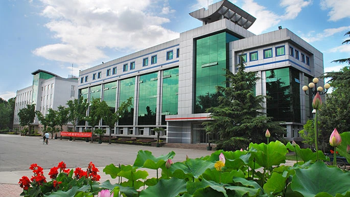
China Lucky was established in 1958 as the Baoding Film Stock Manufacturing Plant, a key project under China’s First Five-Year Plan. In September 2011, it was fully incorporated into the China Aerospace Science and Technology Corporation (CASC).Photovoltaic MaterialsAs China’s largest production base for imaging materials and graphic arts films, and the nation’s most influential manufacturer of optical film materials, Lucky ranks among the world’s top four companies with the capability to produce process-free printing plates. Its products are sold in over 100 countries and regions.Medical MaterialsThe company owns two well-known Chinese trademarks: “Lucky” and “Huaguang.” With a workforce of over 8,600 employees, it operates 12 wholly-owned and holding subsidiaries, 4 directly affiliated units, including two listed companies.Imaging Information Materials | Lucky Tpcw2 Solar Backsheet | Lucky Tpcw1 Transparent Solar Backsheet | pv backsheet manufacturers | solar backsheet suppliers | photo paper bulk | photo paper wholesale | photo paper cost